
Clinical Course of a Shiba Inu With Steroid-Dependent Protein-Losing Enteropathy Managed With Cyclosporine, Dietary Modification, Probiotics, and Adjunctive Huaier-Derived Glycan TPG-1
A generative AI–based explanatory video is available below.
Please note that the AI-generated video may contain interpretations or information not intended in the original case description. For accurate information, please refer to the case details below.
TPG-1 Supplementation Overview
| Dose | Duration |
| 4× the standard feeding amount | 14 days |
| 2× the standard feeding amount | 70 days |
| 3× the standard feeding amount | Approximately 160 days |
TPG-1 supplementation was initiated at four times the standard feeding amount, followed by administration at two times and subsequently three times the standard feeding amount.
Case Information
| Parameter | Details |
| Breed | Shiba Inu |
| Sex | Spayed female |
| Date of birth | July 2, 2017 |
| First presentation | January 22, 2025 (7 years old) |
| Previous diagnosis | Protein-losing enteropathy (PLE) |
Presenting Concerns
The dog had previously been diagnosed with protein-losing enteropathy at another veterinary hospital and had been managed with long-term prednisolone therapy.
Although gastrointestinal signs had been controlled to some extent, attempts to reduce the prednisolone dose were followed by recurrence of soft stools and deterioration of clinical control.
At the same time, reduced muscle condition and orthopedic problems had become clinically important. The dog had a history of orthopedic abnormalities involving the stifle and hip, including cranial cruciate ligament rupture, meniscal injury, and hip luxation.
Dermatologic changes, including scaling, alopecia, and a coarse hair coat, were also present.
These clinical problems were considered potentially related, at least in part, to prolonged glucocorticoid exposure and were negatively affecting the dog's overall condition.
Case Overview
At the first presentation to the reporting hospital, serum albumin concentration was 2.8 g/dL, within the reported reference interval of 2.6–4.0 g/dL.
This suggested that serum albumin was relatively well maintained under the existing treatment regimen. However, attempts to taper prednisolone were associated with recurrence of soft stools, indicating difficulty maintaining clinical control during glucocorticoid reduction.
Findings considered compatible with adverse effects of prolonged glucocorticoid administration, including reduced muscle condition, alopecia, and a coarse hair coat, had also become clinically problematic.
The principal management objective was therefore to maintain control of the underlying enteropathy while progressively reducing systemic glucocorticoid exposure.
The detailed diagnostic workup that originally led to the diagnosis of PLE at the referring hospital was not included in the available case summary. Accordingly, the underlying cause of the protein-losing enteropathy cannot be further characterized from the information presented here.
Treatment Course and Multimodal Management
A multimodal management strategy was introduced with the aim of maintaining gastrointestinal and nutritional stability while permitting gradual reduction of prednisolone.
A. Immunosuppressive and Pharmacologic Management
1. Introduction of cyclosporine
Cyclosporine was introduced as an additional immunosuppressive agent to support disease control during prednisolone tapering.
2. Gradual tapering of prednisolone
Prednisolone was progressively tapered while the dog was managed with cyclosporine and other concurrent interventions.
The long-term goal was complete discontinuation of systemic glucocorticoid therapy if adequate clinical control could be maintained.
B. Dietary and Adjunctive Management
1. Transition to a hydrolyzed diet
The diet was changed to a hydrolyzed diet (z/d®) as part of the management strategy for the chronic enteropathy.
2. Probiotics
Probiotics were introduced on approximately Day 92 after a slight decrease in body weight and serum albumin concentration.
3. TPG-1
TPG-1 was introduced on approximately Day 113 as an adjunctive nutritional intervention.
The initial feeding amount was four times the standard amount for 14 days, followed by twice the standard amount.
On Day 197, the feeding amount was increased to three times the standard amount and supplementation was subsequently continued.
Clinical Course
Before referral, the dog had required long-term prednisolone therapy for management of PLE.
Reduction of the prednisolone dose had repeatedly been associated with recurrence of soft stools, while findings considered compatible with chronic glucocorticoid exposure, including reduced muscle condition, alopecia, and poor coat quality, had become clinically apparent.
At Day 1, cyclosporine was introduced and the diet was changed to a hydrolyzed diet.
Serum albumin remained within or near the reported reference interval during the early treatment period, permitting progressive reduction of prednisolone.
By Day 92, body weight had decreased slightly to 10.60 kg and serum albumin had decreased to 2.4 g/dL. Probiotics were therefore added.
On Day 113, body weight was 10.96 kg and serum albumin was 2.6 g/dL. TPG-1 supplementation was initiated at four times the standard feeding amount for 2 weeks and was subsequently continued at twice the standard feeding amount.
Prednisolone was discontinued on Day 141.
On Day 169, however, an unintended reduction in the administered cyclosporine dose was identified. Prednisolone was therefore reintroduced and the cyclosporine dose was increased.
This episode is important when interpreting the clinical course because it demonstrates that disease management continued to depend on several concurrent treatment adjustments.
On Day 197, TPG-1 was increased to three times the standard feeding amount while prednisolone tapering continued.
Cyclosporine was discontinued on Day 291.
Prednisolone was ultimately discontinued again on Day 354.
At Day 382, serum albumin was 3.0 g/dL, prednisolone remained discontinued, and follow-up was ongoing.
Clinical Timeline
| Day | Date | Body Weight (kg) | Albumin (g/dL) | Prednisolone | Clinical Notes |
| 1 | Jan 22, 2025 | 10.84 | 2.8 | 7.5 mg/head SID | Initial presentation. Cyclosporine initiated and diet changed to z/d®. |
| 22 | Feb 12, 2025 | 10.56 | 3.1 | 3 days on, 1 day off | Albumin remained stable; prednisolone tapering initiated. |
| 50 | Mar 12, 2025 | 10.80 | 2.9 | EOD | Prednisolone further reduced. |
| 71 | Apr 2, 2025 | 10.86 | 2.6 | q96h | Clinical condition remained stable during further tapering. |
| 92 | Apr 23, 2025 | 10.60 | 2.4 | q96h | Slight decreases in body weight and albumin; probiotics added. |
| 113 | May 14, 2025 | 10.96 | 2.6 | q96h | TPG-1 initiated at 4× for 2 weeks, followed by 2×. Cyclosporine dose reduced; an unintended reduction in the administered dose was later recognized. |
| 141 | Jun 11, 2025 | 11.30 | 2.9 | Discontinued | Prednisolone discontinued. |
| 169 | Jul 9, 2025 | 11.50 | 2.5 | q96h | Unintended reduction in cyclosporine dosing identified; prednisolone reintroduced and cyclosporine increased. |
| 197 | Aug 6, 2025 | 11.74 | 2.5 | 5 mg/head q96h | TPG-1 increased to 3×; prednisolone tapering continued. |
| 291 | Nov 8, 2025 | 11.74 | 2.8 | 5 mg/head q96h | Cyclosporine discontinued. |
| 354 | Jan 10, 2026 | 11.92 | 3.0 | Discontinued | Prednisolone discontinued again. |
| 382 | Feb 7, 2026 | 11.30 | 3.0 | Discontinued | Follow-up ongoing. |

Figure 1. Changes in body weight and serum albumin concentration during multimodal treatment.
The treatment timeline includes prednisolone, cyclosporine, clopidogrel, ursodiol, dietary modification with z/d®, probiotics, and adjunctive Huaier-derived glycan TPG-1.
TPG-1 was initiated around Day 113, initially at four times the standard feeding amount, followed by two times and subsequently three times the standard feeding amount.
Because multiple treatments were introduced, discontinued, or dose-adjusted over the same period, the temporal relationship shown in the figure does not establish that TPG-1 caused changes in body weight, serum albumin concentration, or the ability to taper prednisolone.
Clinical Appearance During Follow-up
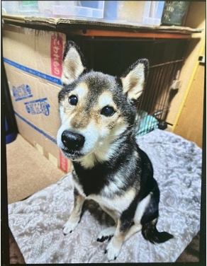
Figure 2A. Day 1.
Generalized alopecia and poor coat quality were observed.


Figure 2B–C. Day 117.
Around the time of TPG-1 initiation, a coarse hair coat and reduced body condition were still evident.

Figure 2D. Day 148.
Approximately 1 month after initiation of TPG-1 supplementation, increased hair coverage was observed along the dorsal and lumbar regions.

Figure 2E. Day 291.
Approximately 6 months after initiation of TPG-1 supplementation, increased hair coverage and an overall improvement in coat appearance were observed.
The photographic changes document improvement in hair coverage over time. However, because prednisolone exposure was also reduced and several other therapeutic and nutritional interventions were administered concurrently, these changes cannot be attributed specifically to TPG-1.
Orthopedic and General Clinical Course
The dog had pre-existing orthopedic disease that had become more clinically problematic during the period of reduced muscle condition.
As the overall medical condition stabilized and glucocorticoid exposure was progressively reduced, episodes of lameness were reported to become less frequent and were subsequently no longer observed during follow-up.
Recovery of body condition and reduced glucocorticoid exposure may have contributed to improved functional stability. However, muscle mass was not quantified using an objective measurement method, and the relative contribution of individual treatments cannot be determined.
At the initial presentation, surgical treatment for the orthopedic disorders had been under consideration.
At the most recent follow-up, the perceived clinical need for orthopedic surgery was reported to be substantially lower, and the owner reported a high level of satisfaction with the dog's overall clinical condition.
No validated quality-of-life assessment tool was used; therefore, improvement in quality of life should be interpreted as an owner- and clinician-reported clinical observation.
Discussion
This case describes the clinical course of a dog with previously diagnosed, steroid-dependent protein-losing enteropathy managed using a multimodal strategy that included cyclosporine, dietary modification, probiotics, progressive glucocorticoid tapering, and adjunctive Huaier-derived glycan TPG-1.
During the observation period, serum albumin remained generally stable, body weight increased from 10.84 kg at initial presentation to a maximum recorded value of 11.92 kg, and prednisolone was ultimately discontinued.
Improvement in hair coverage and coat appearance was also documented photographically during follow-up.
These changes occurred during a period in which several therapeutic interventions were introduced or adjusted.
Importantly, TPG-1 was not the only treatment introduced during this period. Cyclosporine, a hydrolyzed diet, probiotics, and progressive changes in prednisolone dosing were also part of the management strategy.
Prednisolone was first discontinued on Day 141 but had to be reintroduced on Day 169 after an unintended reduction in cyclosporine dosing was identified. Prednisolone was ultimately discontinued again on Day 354.
This sequence demonstrates that the clinical course cannot be interpreted as a simple response to a single intervention.
Accordingly, the independent contribution of TPG-1 to maintenance of serum albumin concentration, body weight gain, improvement in coat condition, or successful glucocorticoid tapering cannot be determined from this single uncontrolled case.
Similarly, although improvement in body condition may have contributed to the reported reduction in orthopedic clinical signs, objective muscle mass measurements were not available.
The improvement in hair coverage may also have been influenced by reduced glucocorticoid exposure, improved nutritional status, changes in gastrointestinal disease control, or other concurrent interventions.
The underlying cause of the dog's PLE was not fully characterized in the available case summary. Therefore, the relevance of this clinical course to other causes of canine PLE remains uncertain.
Nevertheless, this case provides a clinically relevant observation of long-term disease management in which adjunctive TPG-1 was administered throughout a period of successful multimodal treatment and eventual glucocorticoid discontinuation.
The findings should be regarded as hypothesis-generating rather than evidence of a steroid-sparing or therapeutic effect of TPG-1.
Further studies involving clearly characterized PLE populations, standardized treatment protocols, objective clinical outcome measures, and appropriate comparator groups would be required to determine whether adjunctive TPG-1 has a reproducible clinical role in dogs with protein-losing enteropathy.
Conclusion
This case describes a Shiba Inu with previously diagnosed protein-losing enteropathy in which long-term prednisolone therapy had been required to maintain clinical control and was associated with clinically important adverse effects.
A multimodal management strategy incorporating cyclosporine, a hydrolyzed diet, probiotics, progressive prednisolone tapering, and adjunctive Huaier-derived glycan TPG-1 was subsequently implemented.
During follow-up, serum albumin remained generally stable, body weight increased, hair coverage improved, and prednisolone was ultimately discontinued.
However, because multiple therapeutic and nutritional interventions were administered concurrently and adjusted throughout the observation period, these outcomes cannot be attributed specifically to TPG-1.
In particular, this single case does not establish that TPG-1 has a glucocorticoid-sparing effect, improves muscle mass, restores coat quality, or directly controls PLE.
The clinical course should therefore be interpreted as a preliminary observation supporting further investigation of TPG-1 as a potential adjunct within multimodal management of canine protein-losing enteropathy.