
Case provided by Professor Masaharu Hisasue, Azabu University Veterinary Teaching Hospital
Case Information
A 10-year-6-month-old spayed female Toy Poodle weighing 5.62 kg (body condition score, 2.5/5) had a history of immune-mediated polyarthritis and immune-mediated thrombocytopenia (IMTP). These conditions had been managed with prednisolone and mycophenolate mofetil and were reported to be clinically well controlled at the time of evaluation.
On physical examination, the body temperature was 38.8°C, heart rate was 160 beats/min, and respiratory rate was 36 breaths/min. Hematologic findings included a white blood cell count of 7,460/μL, neutrophil count of 5,240/μL, red blood cell count of 6.92 × 10⁶/μL, hemoglobin concentration of 16.6 g/dL, packed cell volume of 48%, platelet count of 482 × 10³/μL, and reticulocyte count of 63 × 10³/μL. The serum C-reactive protein concentration was 0.9 mg/dL.
At that time, the platelet count was within the reference range, and there was no hematologic evidence of active thrombocytopenia.
Diagnostic Evaluation
The dog was re-evaluated because of worsening halitosis. Periodontal disease was initially suspected, and dental scaling was performed at the primary care veterinary clinic. During the procedure, a mass was identified at the base of the tongue.
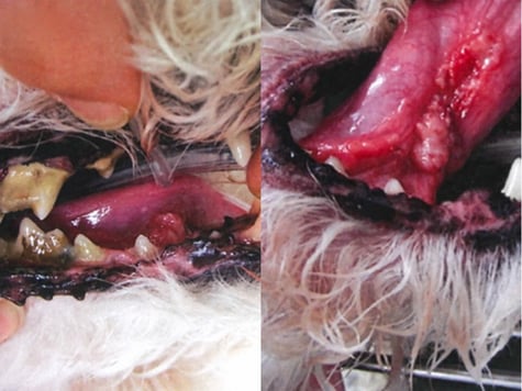
Figure 1. Gross oral findings at the time of dental scaling.
The mass was partially excised, and histopathologic examination resulted in a diagnosis of squamous cell carcinoma (SCC).
On subsequent evaluation by the surgical oncology service, induration of the tongue was detected on palpation. Diagnostic imaging did not identify enlargement of the mandibular or medial retropharyngeal lymph nodes, and cytologic evaluation of the regional lymph nodes did not reveal evidence of metastasis. No evidence of distant metastasis was identified on the examinations performed at that time.
However, complete local tumor control could not be assumed after partial excision, and the possibility of residual local disease, subsequent local recurrence, or microscopic vascular invasion could not be excluded. Complete surgical excision, with consideration of adjunctive radiotherapy, was therefore presented as the preferred definitive treatment approach.
Treatment and Clinical Course
The owner elected not to pursue definitive surgery or radiotherapy. Given the dog's age and concerns regarding the overall burden associated with more intensive treatment, the owner's preference was to prioritize maintenance of quality of life and voluntary food intake while minimizing treatment-related burden.
TPG-1 was subsequently initiated at twice the usual product dose as an adjunctive intervention. TPG-1 has been investigated for potential immunomodulatory activity; however, its clinical efficacy for the treatment of canine lingual SCC has not been established. Its use in this case was therefore considered adjunctive and was not intended to replace standard local oncologic treatment.
At the time of diagnosis, the prognosis was considered guarded. During subsequent follow-up, no clinically apparent rapid enlargement of the lingual lesion was observed. At approximately 6 months after initiation of adjunctive TPG-1, no marked gross change in the appearance of the tongue lesion was noted.
During this period, no clinically apparent distant metastasis or rapid local progression was identified. The dog's general condition remained stable, and voluntary food intake was maintained.
Because no marked clinical progression of the tumor had been observed during follow-up, continued administration of TPG-1 was proposed. TPG-1 was subsequently discontinued on Day 397 of the reported clinical course.
The dog's clinical condition remained manageable thereafter, and the dog died on Day 566 following the development of an intracranial hemorrhage. Recurrence of IMTP was considered a possible underlying cause of the hemorrhage based on the clinical course; however, a definitive causal relationship between recurrent IMTP and the intracranial hemorrhage was not established.
No rapid progression of the lingual SCC or clinically apparent distant metastasis had been documented before death.
Discussion
Published outcomes for canine lingual SCC vary considerably according to tumor extent, disease stage, treatment, and patient population. Historical reports summarized in the source material describe median survival times of approximately 120–216 days in dogs with lingual SCC, with 1-year survival rates of 25% or less in some reported populations. Longer survival has also been described in individual dogs treated with radiotherapy or multimodal therapy.
The reported clinical course in the present case extended to 566 days. During much of this period, no clinically apparent rapid local progression or distant metastasis was documented, and voluntary food intake and general condition were maintained.
These observations should not, however, be directly compared with historical survival estimates. Differences in tumor size and extent, disease stage, surgical treatment, treatment protocols, patient characteristics, and follow-up methods may substantially influence outcome.
In the present case, the lingual mass had initially been partially excised, whereas definitive surgical excision and radiotherapy were recommended but were not pursued. TPG-1 was administered as an adjunctive intervention, and a relatively prolonged period of clinical stability was subsequently observed.
Importantly, however, this temporal association does not establish an antitumor effect of TPG-1. Because this report describes a single uncontrolled case, it is not possible to determine whether the observed clinical course was related to TPG-1 administration. The initial partial excision, the individual biological behavior of the tumor, the extent of disease at diagnosis, host-related factors, concomitant medications, and other unmeasured variables may also have influenced the outcome.
TPG-1 was discontinued on Day 397, and the dog remained alive until Day 566. Therefore, the overall survival duration cannot be directly attributed to continued TPG-1 administration.
In addition, the absence of clinically apparent progression does not necessarily indicate complete tumor control. Serial standardized measurements of tumor dimensions and repeated advanced imaging were not documented throughout the entire observation period; therefore, subclinical local progression or metastatic disease cannot be excluded.
Accordingly, the present case should be interpreted as a report of prolonged clinical stability in a dog with lingual SCC that received adjunctive TPG-1 after partial excision, rather than as evidence of therapeutic efficacy of TPG-1.
Further accumulation of well-documented cases, standardized assessment of tumor burden and response, systematic evaluation of quality of life, and preferably controlled prospective studies would be required to determine whether adjunctive TPG-1 has any clinically meaningful effect on tumor progression, quality of life, or survival in dogs with squamous cell carcinoma.